Stone Treatments
Kidney stones can be treated in several ways, depending on the size and location of the stone and the individual's overall health. Treatment options may include:
- Medications to help assist in relaxing the ureter to aid stone passage
- Pain relief during the stone passage
- Laser Stone Surgery
- Percutaneous Nephrolithotomy (PCNL)
- Extracorporeal Shock Wave Lithotripsy (ESWL)
- Ureteroscopy
Flexible Pyeloscopy Laser Stone Surgery
Minimally Invasive Surgery (MIS) uses the latest advanced technology to treat pain and other symptoms caused by various urological disorders.
Holmium Laser Stone Surgery using Flexible Pyeloscopy (or Retrograde IntraRenal Surgery- RIRS) is where a thin, lighted telescope is introduced into the kidney from outside the patient without the need for any incision (see diagram).
Minimally Invasive Pyeloscopy
The diameter of the instrument is 2.5mm and allows visualisation of the entire kidney and the ureter due to the flexible nature of the scope.
It contains a small instrument port that introduces a holmium laser fibre (0.2 mm diameter) to efficiently fragment stones and micro-baskets (less than 1mm wide) to retrieve stone fragments.
Kidney stones up to 1.5cm in size can be treated using this approach.
Advantages of Flexible Pyeloscopy
Benefits of Flexible Pyeloscopy over traditional methods include the following:
- Treatment without scars
- A high degree of success
- It can be performed as day surgery
- Less risk of infection
- Minimal postoperative pain
- Quicker recovery
- Quicker return to work and normal activities
Disadvantages of Flexible Pyeloscopy
Compared with shock wave lithotripsy:
- A general anaesthetic is required, and
- Small risk of damage to the ureter (0.5%).
Before Flexible Pyeloscopy
Preparation for the procedure is performed under general anaesthesia; you should have nothing to eat or drink for 6 hours before treatment.
You will meet your anaesthetist, who will take a thorough medical history before surgery. This person will be responsible for your safety while under general anaesthesia.
Regular medications can be taken with a sip of water except for blood thinning agents (e.g. warfarin, aspirin, clopidogrel) or non-steroidal anti-inflammatories, which may need to be stopped beforehand. Your doctor will advise you about your specific circumstances.
A midstream urine (MSU) test is required to ensure the urine is sterile before treatment.
During Flexible Pyeloscopy
The procedure will usually take 60 to 90 minutes and involves putting a flexible telescope into the drainage tube of the kidney and fragmenting the stone(s) with a holmium laser.
A temporary double J ureteral stent may be left in place for a short period to ensure the kidney drains without risk of blockage.
After Flexible Pyeloscopy
It is normal to feel the need to pass urine frequently and notice blood in the urine following surgery.
This will settle over the ensuing days. An oral over-the-counter medication called the Ural can reduce the stinging sensation during urination.
You will sometimes have a temporary ureteral stent (see stent info sheet) following surgery, allowing the stone fragments to drain unimpeded.
The stent may be attached to a string tether coming out from the urethra, which allows easy removal in the doctor’s office. Care must be taken to avoid accidentally dislodging the stent by letting it catch up on your underwear.
Care Plan After Flexible Pyeloscopy
You will be advised of the necessary follow-up arrangements after surgery. A script for oral antibiotics will need to be taken for five days to prevent infection.
You must drink at least eight glasses of water daily (2.5L/day). Simple analgesics such as Panadol and Nurofen are usually all required; occasionally, stronger medication (e.g. Panadeine Forte) may be necessary.
You will not be able to drive for at least 24 hours after surgery as you have had a general anaesthetic.
Flexible Pyeloscopy Risks
This is generally considered a very safe operation. Surgical risks include
- infection,
- minor bleeding, and
- perforation of the ureter (1 in 200).
Percutaneous Nephrolithotomy (PCNL)
Percutaneous Nephrolithotomy (PCNL) is a minimally invasive surgical procedure to remove large kidney stones. It involves making a small incision in the back, usually in the area of the kidney where the stone is located, and using instruments to remove the stone.
PCNL is typically recommended for individuals with large kidney stones that cannot be treated with other methods such as ESWL, ureteroscopy or observation. It is also suitable for individuals with a history of recurrent kidney stones or stones in the lower pole of the kidney that are not amenable to other treatments.
Types of Percutaneous Nephrolithotomy
- Mini PCNL (Percutaneous Nephrolithotomy) is a newly developed technique for the percutaneous removal of renal stones.
- Supine percutaneous nephrolithotomy (PCNL) is a novel, safe approach to removing kidney stones. The procedure is performed with the patient in a supine position.
Benefits of Percutaneous Nephrolithotomy
Some of the benefits of PCNL include the following:
- High success rate: PCNL has a high success rate in removing large kidney stones.
- Minimally invasive: The procedure is minimally invasive and does not require a large incision.
- Faster recovery: Recovery time is generally quicker than traditional open surgery.
- Preservation of kidney function: By removing the stones before they cause damage to the kidneys, PCNL helps to preserve kidney function
Before Percutaneous Nephrolithotomy
Before the procedure, the patient will need to fast for 6 hours. They will also be given general anaesthesia or spinal anaesthesia. The patient will be placed in a prone position on an x-ray table.
During Percutaneous Nephrolithotomy
A small incision, typically around 1-2 cm, is made in the back, near the kidney where the stone is located. A nephroscope (a tiny telescope) is inserted through the incision and into the kidney to find the stone.
The stone is then fragmented using a laser or other instrument and removed. A small tube may be left in the kidney for a few days to prevent bleeding and infection.
Percutaneous Nephrolithotomy Recovery
After the procedure, the patient will be monitored in the recovery room and then moved to a hospital room for some time, usually overnight. The patient may need to remain on bed rest for a day or two. Drainage tubes may be left in place for a few days to help prevent infection and bleeding.
Care Plan for Percutaneous Nephrolithotomy
After the procedure, the patient will be instructed to drink lots of water and maintain a low-salt diet to help prevent the formation of new stones. The patient will also be advised to avoid heavy lifting and strenuous activities to allow the incision to heal. Regular follow-up appointments with the healthcare provider will be scheduled to ensure the recovery is going well and to check for any complications.
Percutaneous Nephrolithotomy Risks
The risks of PCNL include bleeding, infection, injury to surrounding organs, and the formation of new stones. It's important to discuss the risks and benefits of PCNL with your healthcare provider before the procedure to determine if it's the best option for you.
Extracorporeal Shock Wave Lithotripsy (ESWL)
What is Extracorporeal Shock Wave Lithotripsy?
Extracorporeal Shock Wave Lithotripsy (ESWL) is a non-invasive procedure used to break up kidney stones into small pieces that can be passed through the urinary tract. It involves using high-energy shock waves directed at the stone from outside the body.
ESWL is typically recommended for individuals with small to medium-sized kidney stones that are located in the upper or middle portion of the ureter. It may also be recommended for individuals with recurrent kidney stones or stones that cannot be treated with other methods, such as ureteroscopy or observation.
Benefits of ESWL
Benefits of ESWL include
- High success rate: ESWL has a high success rate in breaking up small to medium-sized kidney stones.
- Non-invasive: The procedure is non-invasive and does not require a large incision.
- Faster recovery: Recovery time is generally quicker than with invasive procedures.
- Preservation of kidney function: By breaking the stones before they cause damage to the kidneys, ESWL helps to preserve kidney function
Before ESWL
The patient will be given general or spinal anaesthesia and positioned on a table with the stone in the targeted area. The patient may be asked to drink large amounts of water before the procedure to help locate the stone with X-ray or ultrasound images.
During ESWL Procedure
The patient will lie on a water-filled cushion, and the machine will direct high-energy shock waves at the stone from outside the body, breaking it into smaller pieces. The treatment session typically lasts for about 45-60 minutes.
ESWL Recovery
After the procedure, the patient will be monitored in the recovery room and discharged on the same day. The patient may need to remain on bed rest for a day or two.
Care Plan for ESWL
After the procedure, the patient will be instructed to drink lots of water and maintain a low-salt diet to help prevent the formation of new stones and help the smaller pieces pass through the urinary tract. The patient will also be advised to avoid heavy lifting and strenuous activities to allow the body to pass the smaller stones.
Regular follow-up appointments with the healthcare provider will be scheduled to ensure the recovery is going well and to check for any complications.
ESWL Risks
The risks of ESWL include bleeding, infection, injury to surrounding organs, incomplete clearance and the formation of new stones. It's important to discuss the risks and benefits of ESWL with your healthcare provider before the procedure to determine if it's the best option for you.
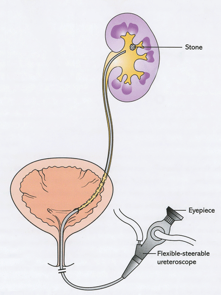
Ureteroscopy
What is Ureteroscopy?
Ureteroscopy is a procedure used to diagnose and treat kidney stones. It involves using a small telescope (ureteroscope) inserted through the urethra and into the bladder to reach the ureter and the kidney, where the stone is located.
Ureteroscopy is typically recommended for individuals with small to medium-sized kidney stones that are located in the upper or middle portion of the ureter. It may also be recommended for individuals with recurrent kidney stones or stones that cannot be treated with other methods, such as ESWL or observation.
Benefits of Ureteroscopy
Benefits of ureteroscopy include:
- High success rate: Ureteroscopy has a high success rate in removing small to medium-sized kidney stones.
- Minimally invasive: The procedure is minimally invasive and does not require a large incision.
- Faster recovery: Recovery time is generally quicker than with traditional open surgery.
- Preservation of kidney function: By removing the stones before they cause damage to the kidneys, ureteroscopy helps to preserve kidney function
Ureteroscopy Procedure
During the procedure, your doctor:
- Inserts the tip of the ureteroscope through the urethral opening (opening of the urinary bladder to the outside)
- Advances it up the urinary bladder and into the ureter (kidney tubules empty urine into the bladder).
- A sterile liquid is passed through the ureteroscope to fill and stretch the bladder and ureter. This helps your doctor to view the interior wall clearly, locate the stone and diagnose any abnormalities.
- To remove the stone, your doctor may insert a thin wire with a tiny basket attached to its end through the ureteroscope.
- The basket grabs and removes the kidney stone.
- Your doctor may sometimes break a large stone with the help of a laser emitted through a flexible fibre passed through the ureteroscope.
During a ureteroscopy, no incisions are made. It is an outpatient procedure performed under spinal or general anaesthesia.
The complete procedure takes about 30-60 minutes.
After Ureteroscopy Procedure
After the procedure, you may feel a mild burning sensation during urination.
You will be advised to drink plenty of water to relieve postoperative discomfort. Your doctor may prescribe antibiotics for a few days to prevent infection.
Ureteroscopy Risks
The risks of ureteroscopy include bleeding, infection, injury to surrounding organs, and the formation of new stones. It's important to discuss the risks and benefits of ureteroscopy with your healthcare provider before the procedure to determine if it's the best option for you.
Kidney Stone Treatments Prognosis
The prognosis for kidney stone treatment depends on the size and location of the stone, as well as the individual's overall health and medical history.
Treatments such as drinking plenty of water, taking pain relievers, and undergoing minimally invasive procedures such as ESWL, ureteroscopy, or PCNL have a high success rate and a relatively short recovery time.
What if Kidney Stone Treatment is Delayed?
If kidney stone treatment is delayed, the stone may continue to cause pain and discomfort, leading to complications such as infection, blockage of the urinary tract, or damage to the kidney.
Additionally, the longer the stone remains in the urinary tract, the greater the chance it will grow larger, making it more difficult to remove. You must seek medical treatment as soon as possible if you suspect you have a kidney stone.